
Several conditions under one name
Cardiomyopathy is a family of conditions rather than a single disease. The main types differ in what the muscle does wrong:
- Dilated cardiomyopathy: the main pumping chamber enlarges and the muscle weakens, so each beat moves less blood
- Hypertrophic cardiomyopathy: the muscle thickens, often unevenly, making the chamber stiff and sometimes obstructing blood flow
- Restrictive cardiomyopathy: the muscle stiffens without thickening much, so the heart cannot fill properly
- Arrhythmogenic cardiomyopathy: muscle is gradually replaced by scar and fatty tissue, raising the risk of rhythm problems
- Left ventricular noncompaction: the muscle wall has a spongy, deeply grooved inner layer that formed differently before birth
Dilated and hypertrophic forms are by far the most common in children. Some children show features of more than one type, and the label can change as more information comes in. The type matters because it shapes which tests come next and which treatments make sense.
Where it comes from
In many children, no single cause is ever found. In others, the cause becomes clear with time and testing.
Genetic changes are a major source, particularly for hypertrophic cardiomyopathy. These are often inherited from a parent who may have no symptoms at all, which is why a diagnosis in a child frequently leads to screening of parents and siblings. Our earlier post on family history screening explains when relatives should be checked.
Viral infections can inflame the heart muscle, a condition called myocarditis. Most children recover fully, but in some the inflammation leaves the muscle weakened, resulting in dilated cardiomyopathy. That is why a child who seems unusually breathless or exhausted in the weeks after an ordinary virus, out of proportion to the illness, is sometimes sent for an echo. Most such scans are normal. The few that are not are found while there is still room to treat.
Other causes include neuromuscular conditions such as Duchenne muscular dystrophy, inherited metabolic disorders, certain chemotherapy medicines used in childhood cancer, and long periods of very fast heart rhythm, which can tire the muscle until the rhythm is controlled. That last cause is one of the few that is often fully reversible.
How it shows itself, or does not
Symptoms depend on the type and how advanced it is. Some children have none, and the diagnosis comes from a murmur, an abnormal ECG at a sports screening, or family screening after a relative is diagnosed.
When symptoms are present, they tend to look like a heart that cannot keep up with demand. In babies, that means fast breathing, sweating and tiring during feeds, and slow weight gain. In older children, it can mean tiring more quickly than friends, breathlessness with activity, a persistent cough, swelling around the eyes or ankles, tummy pain or poor appetite.
The tiredness is the hardest sign to read, because children are tired for many reasons. Our post on children who tire faster covers the difference between ordinary fatigue and the kind that deserves a heart check.
Some signs need urgent attention whatever the background: fainting, especially during exercise; chest pain with exertion; a racing heartbeat with dizziness; or breathlessness at rest. In hypertrophic cardiomyopathy in particular, fainting during exercise can be a warning of a dangerous rhythm.
Building the full picture
The echocardiogram is the central test. It measures the size of each chamber, the thickness of the walls, how well the muscle squeezes and relaxes, and whether blood flow is obstructed.
An ECG looks for rhythm problems and patterns typical of particular cardiomyopathies. A Holter monitor records the rhythm over a day or more. A cardiac MRI can show scar tissue and inflammation in the muscle that ultrasound cannot see, which helps with both diagnosis and risk assessment.
At our Dubai Healthcare City clinic, the echo and ECG are usually done at the first visit, so the family leaves with a clearer picture the same day. Further tests are then scheduled according to what those first results show, rather than all at once.
Genetic testing is now a routine part of the workup for many children. A confirmed genetic cause can clarify the type, guide which relatives need screening, and sometimes point toward specific treatments.
When a genetic form is found or suspected, parents and brothers and sisters are usually offered an ECG and echocardiogram. Children in the family who test normal may still be rechecked every few years through adolescence, because some forms, hypertrophic cardiomyopathy especially, can appear during growth spurts rather than at birth.
For children who want to play sport, especially those with hypertrophic cardiomyopathy, a careful sudden cardiac risk assessment helps the cardiologist and family agree on which activities are reasonable. Guidance in this area has become more individualized in recent years, with less blanket restriction and more shared decision-making.
Living with the diagnosis
Treatment aims to ease the work of the heart, control symptoms, and prevent complications. Depending on the type, that may include medicines that help a weak heart pump more efficiently or a stiff heart relax, medicines that control rhythm, and diuretics to clear excess fluid.
Some children with a high risk of dangerous rhythms are offered an implantable cardioverter-defibrillator (ICD), a small device that can detect and correct a life-threatening rhythm. For a small number with severe, progressive heart failure, referral to a specialist center for advanced therapies, including heart transplantation, becomes part of the plan.
Everyday life usually continues with some adjustments. Children with cardiomyopathy are generally advised to keep up with routine vaccinations, including the yearly flu vaccine, since a bad respiratory infection places extra strain on a weakened heart. On medicines such as diuretics, a stomach bug with vomiting or diarrhea can cause dehydration more quickly than usual, so families are given a plan for sick days. Schools need a copy of the cardiologist’s activity advice, and a named contact who knows what to do if the child feels unwell.
Outcomes vary widely. A child whose dilated cardiomyopathy followed myocarditis may recover substantial heart function over months. A child with mild hypertrophic cardiomyopathy may need little more than regular monitoring for years. Others need intensive treatment. The cardiologist’s role is to work out which path a particular child is on, and to keep reassessing as the child grows.
The seven-year-old whose echo report opened this post is a good example of the first path. His story turned toward recovery: rest, medication and repeat scans over several months, and a heart that gradually returned toward normal. Not every child’s course looks like his. Every child’s course is better understood with careful follow-up.
Practical points for the months ahead
Families leaving a first cardiomyopathy appointment often have more questions than they can hold. A few practical steps help.
Keep copies of every echo and MRI report, and bring them to each visit; the trend over time often says more than any single result. Ask which symptoms should prompt a same-day call and which can wait for the next appointment. Ask whether parents and siblings should be screened, and when. Share the cardiologist’s written advice with the school, particularly about PE and sport. Keep a running list of questions on a phone between visits, since the ones that matter most tend to surface at home rather than in the clinic. And if a diagnosis feels unclear, asking for the images to be reviewed by a second cardiologist is a normal request, not a vote of no confidence.
Follow-up at our Dubai Healthcare City clinic usually combines repeat echocardiograms with clinical review at intervals set by how stable the heart is. Families in Abu Dhabi and Al Ain can have much of that follow-up nearer home, with the Dubai team involved where specialized imaging or decisions are needed. Our Dubai cardiology team can be reached through the KidsHeart contact page to arrange an assessment or a second look at an echo report that raised a question.
Consultant, Fetal & Pediatric Cardiologist – American Board
Assistant professor, Columbia University, New York
Pediatric Cardiac Surgeon – American Board 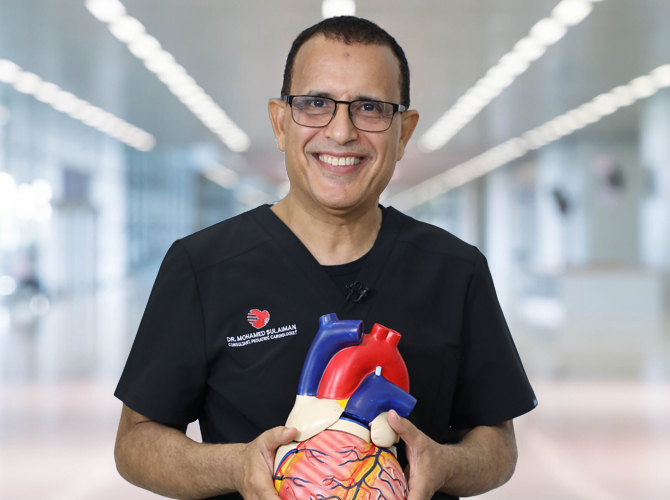
Dr. Mohamed Sulaiman

Dr. Emile A. Bacha